ライフサイエンス関連資料
ダウンロード: ライフサイエンス
-
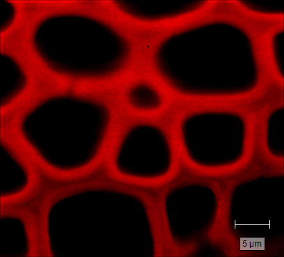
Brochure: Biological analysis using Raman spectroscopy and imaging [en]
The domain of biological research is shaped by our ability to peer into the world of the small. Simply seeing microscopic biological samples is useful, but by utilising Raman spectroscopy we can surpass sight into the molecular realm… and beyond! Download this brochure to discover the wealth of biological applications made possible by Renishaw's Raman systems.
-
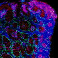
アプリケーションノート: inVia コンフォーカルラマンマイクロスコー プのレドックス生物学への応用
ヘムタンパク質の存在を検出できるラマン分光は、単離や染色を行う必要がなく、レドックス生物学の研究に理想的です。ヘムタンパク 質のレドックスは、酸素の運搬と貯蓄、電子の伝達、フリーラジカルの除去といったタンパク質の機能性に密接に関わっています。そのた め、生体システム内のレドックス状態の解明にラマン分光を使用することで、レドックスダイナミクスおよび健康調整と疾病に対する影響 を研究することができます。
-
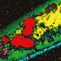
アプリケーションノート: バイオアプリケーション用ラマンイメージング。 染色もラベルも不要
ラマン分光は、多彩な情報が得られるラベルフリーの非侵襲的なイメージング技術で、ライフサイエンス研究に理想的です。レーザー光 の拡散を使用して、分析領域の各ポイントにおける化学的な指紋を取得し、サンプル内の分子を特定します。
-
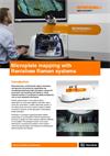
Product note: Microplate mapping with Renishaw Raman system's [en]
Renishaw’s microplate mapping package enables researchers to use Renishaw’s Raman spectroscopy products to rapidly and easily analyse material contained in microplates.
ダウンロード: ライフサイエンス (細胞)
-
Application note: Classification of brain glioma tumours using the Renishaw Biological Analyser [en]
Demonstrating discrimination between diseased and healthy brain tissue using the Renishaw RA816 Biological Analyser.
-
アプリケーションノート: inVia™ コンフォーカルラマンマイクロスコープによる細胞イメージの取得
ラマンイメージにより、細胞の生化学的組成を変容させることなく、細胞種を判別して細胞下構造を解明することができるようになりました。このラベルフリーの非接触、非破壊分析技術は、サンプルの準備がほとんど必要ないため、生細胞の分析とイメージングに理想的です。
-
アプリケーションノート: アプリケーションノート : inVia™ コンフォーカルラマンマイクロスコープによる表面増強ラマンスペクトル(SERS)イメージの取得
ラマンイメージは、各種化学種の分子組成、構造、分布の理解に役立つ強力な研究ツールです。SERS では、ナノシルバー/ゴールドコロイドや粗面化金属基板を使用して、吸収される分子のラマン散乱強度を増幅できます。これにより分析感度や特異性を向上することができます。
-
Application note: Raman imaging to reveal components and metabolites in wood cells and tissue [en]
Analysing Scots pine wood using the inVia™ confocal Raman microscope, to reveal high-resolution details of structure and chemical composition.
-
Portrait of a dying cell [en]
The December 2015 issue of 'The Pathologist' featured an article describing how Raman spectroscopy is a non-invasive way of obtaining morphological and chemical information about cells that may lead to better cancer research.
ダウンロード: ライフサイエンス (組織)
-
Application note: Monitoring transdermal drug delivery in skin using the RA816 Biological Analyser [en]
Confirming the presence of a topical compound in the epidermis and reticular dermis with high specificity and sensitivity.
-
Application note: Classification of brain glioma tumours using the Renishaw Biological Analyser [en]
Demonstrating discrimination between diseased and healthy brain tissue using the Renishaw RA816 Biological Analyser.
-
アプリケーションノート: inVia ラマンマイクロスコープによる組織 イメージの取得
組織のラマンイメージは、ラベリングを施すことなく、組織内の分子組成と各化学種の分布を高い空間分解能で同時に調べることができるユニークな手段です。
-
Application note: Raman imaging to reveal components and metabolites in wood cells and tissue [en]
Analysing Scots pine wood using the inVia™ confocal Raman microscope, to reveal high-resolution details of structure and chemical composition.
-
Product note: Rayleigh imaging using the inVia™ confocal Raman microscope [en]
Product note detailing how you can perform Rayleigh imaging on the inVia confocal Raman microscope.
-
Application note: Virsa Raman analyser for in vivo studies [en]
The Virsa™ Raman analyser is ideal for developing clinical applications of Raman spectroscopy. It is available with a clinically-ready fibre-optic probe developed in partnership with EmVision, leaders in medical fibre probes.
ダウンロード: ライフサイエンス (植物生物学)
-
Application note: Raman imaging to reveal components and metabolites in wood cells and tissue [en]
Analysing Scots pine wood using the inVia™ confocal Raman microscope, to reveal high-resolution details of structure and chemical composition.
-
Application note: Raman imaging of plant metabolite crystals [en]
Raman imaging is an ideal technique for studying plant tissue in a non-destructive and non-invasive way. Raman images spatially resolve chemical and structural information, and can be acquired in the presence of water and in vivo. Here the spatial distribution of plant metabolites and tissue structure are revealed using the Renishaw inVia™ Qontor® confocal Raman microscope.
ウェビナー
ウェビナー - 「レドックス生物学のための共鳴ラマン分光法」もご覧ください。
活用事例
レニショーでは、次のような各種活用事例集を作成しています。
詳細については、下のボタンからレニショーまでお問い合わせください。その際、目的の文書の参照番号をお伝えください。
文書の参照番号 | 説明 |
AS013 | 神経膠腫細胞の 3D イメージ。 inVia と 3D イメージにより細胞の情報を解明します。 |
AS014 | ラットの脊椎組織の 3D イメージ。 inVia と 3D イメージにより組織の情報を解明します。この例では、ラットの脊柱組織の構成要素を調べています。 |
AS016 | 細胞小器官の 3D イメージ。 inVia と 3D イメージにより細胞の情報を解明します。この例では、神経膠腫細胞の細胞小器官のサイズと位置を調べています。 |
AS017 | 精子のイメージ。 inVia により、有機体の解剖学的構造に関する情報を解明します。この例では、精子の解剖学的構造を調べています。 |
AS024 | 神経膠腫細胞の AFM イメージと 3D ラマンイメージ。 原子間力顕微鏡 (AFM) イメージと inVia 3D ラマンイメージにより神経膠腫細胞の組成に関する詳細情報を解明します。 |
AS025 | 細胞小器官の蛍光イメージと 3D ラマンイメージ。 共焦点 3D ラマンイメージと蛍光イメージにより神経膠腫細胞の組成に関する詳細情報を解明します。 |
AS031 | ラベリングを施すことなく胚性幹細胞/癌腫の細胞株を区別。 この例では、StreamLine™ イメージと主成分分析を使用して、ラベリングを施していない胚性幹細胞 (ESC) と胚性癌腫細胞 (ECC) の細胞株を区別しました。 |
AS034 | 精子のラマンイメージ。 inVia により正常な細胞と異常な細胞の解剖学的、化学的情報を解明します。この例では、表現型正常の精子と表現型異常の精子の解剖学的構造を調べています。 |
AS036 | 植物細胞壁のラマンイメージ。 StreamLineHR イメージを使用して植物細胞壁とシリンギルリグニンの分布と相対厚を解明します。 |
AS037 | メラノーマ工学モデルのラマンイメージ。 工学 3D 合成を使用して、メラノーマなどの腫瘍の生化学的組成を調べたり、疾患モデルに対する薬の影響を調べます。通常、腫瘍のサイズ、化学情報、隣接組織への侵食と化学的変化を評価します。 |
AS038 | ラマンの生細胞イメージ。 非侵襲式生細胞イメージ。 |
AS053 | ラマンイメージによりアポトーシス細胞の形態的、生体分子的特徴を解明。 共焦点 StreamLineHR イメージを使用してアポトーシス細胞の形態的特徴を解明します。 |