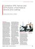
ハイブリッドラマンシステム
他社製分析システムと組み合わせることで、レニショーラマンマイクロスコープのパフォーマンスが向上します。
装置間でサンプルを移動することなく、複数の技術で分析を行うことにより、効率を高められます。
また、レニショーの相関性顕微システムを使用することで、両方の技術で同時に同じ位置を分析できます。
問合せ
SPM/AFM: ナノレベルの分解能
inVia™ ラマンマイクロスコープに、AFM (原子間力顕微鏡) などの走査型プローブ顕微鏡 (SPM) を統合することで、物質の構造および化学特性を調べることができます。さらに、チップ増強ラマン分光測定 (TERS) によりナノメートル単位の化学的分解能を追加し、力学的特性などの補足情報を明らかにできます。
ナノインデンテーション: 機械特定の測定
ナノインデンテーション測定に inVia ラマンマイクロスコープの機能を応用し、機械特性およびトライボロジ特性と、結晶化度、結晶多形性、位相、応力などの化学情報を直接相関させます。
『Microscopy and Analysis』誌の記事をご覧ください。
-
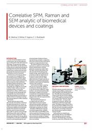 Correlative SPM Raman and SEM analysis of biomedical devices and coatings [en]
A variety of medical implants and microelectrode arrays for electrophysiology are fabricated in thin film and micro technology. To guarantee the quality, proper functionality and safe operation of these devices, analytical techniques to investigate the structure and chemical composition of surfaces and interfaces during the fabrication process and for final quality control are essential.