Renishaw to present a paper and lead a workshop at MMC 2015
MMC 2015, incorporating EMAG 2015, is hosted by the Royal Microscopical Society and takes place from 29th June to 2nd July 2015, at the Manchester Central Convention Complex, Manchester, UK.
During this exhibition and conference, Applications Scientist, Dr Katherine Lau, will lead a workshop entitled “Raman confocal microscopic imaging – a powerful label-free tool for life sciences research.” Taking place on Tuesday 30th June at 3:30 p.m., in area 3, it is a fantastic opportunity for delegates to gain a greater understanding of how Raman imaging can simultaneously delineate the chemical and morphological information of biological samples.
On Wednesday 1st July at 10:45 a.m., Katherine will also present a paper during the Imaging Cancer session. The paper “Revealing multiple cellular changes in autophagy-induced and apoptotic MG-63 osteosarcoma cells using label-free Raman confocal microscopic imaging” is co-authored by scientists, Bhagavathi Ramamurthy and Frederick Coffman from the Center for Biophysical Pathology at Rutgers New Jersey Medical School. Katherine's presentation will demonstrate the use of Raman microscopic imaging (RMI), a label-free information-rich technique, to reveal morphological and chemical changes associated with autophagy and apoptosis in cancer cells.
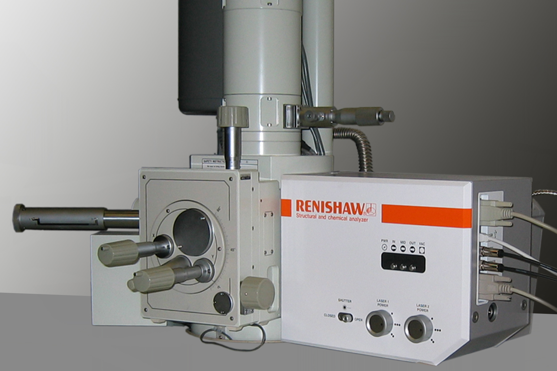
Visitors to stand 504, will be able to discuss Raman applications, as well as Renishaw's range of Raman products, including combined techniques, such as AFM-Raman and SEM-Raman. When asked what Renishaw is showing on the stand, Martin Davies, UK Sales Manager, said: “Come and speak to our team about how Raman, coupled with a scanning electron microscope (SEM), gives you chemical structure information from your samples. Raman complements energy-dispersive x-ray spectroscopy (EDS) elemental analysis, giving clear and precise detail of sample defects, stress, polymorph differentiation, crystallographic structure and film thickness. We would be happy to discuss your research needs and how Renishaw's Structural and Chemical Analyser can help.”
For further details of Renishaw's Structural and Chemical Analyser and its application to study a broad variety of samples, please visit www.renishaw.com/SEMRaman